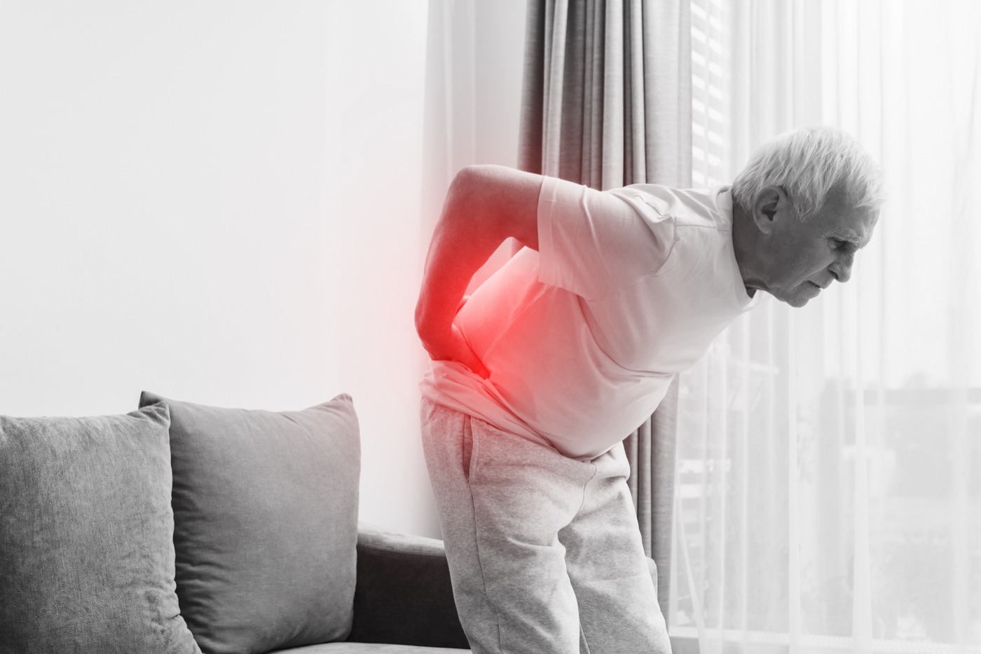
Between the vertebrae of your spine sit intervertebral discs — cartilaginous shock absorbers that distribute load and enable movement. Each disc has a tough outer ring (annulus fibrosus) and a gel-like inner core (nucleus pulposus). In normal aging, discs gradually dehydrate and degenerate. In a car accident, the sudden, forceful spinal loading can tear the outer ring and force the inner material outward — compressing nerve roots, causing intense radiculopathy, and producing symptoms that can persist for months or years.
Herniated discs are among the most serious and consequential car accident injuries. They're also among the most documentable — unlike purely soft tissue injuries, disc herniation is visible on MRI with high specificity. That objective documentation is what connects the severity of the symptoms to the injury in the medical record.
How car accidents cause disc herniation
Different crash types load the spine differently, but the underlying mechanism is similar: sudden, unexpected spinal loading at forces and speeds that exceed normal physiological tolerance.
In rear-end collisions — the most common cause of cervical disc herniation — the struck vehicle accelerates forward while the occupant's head lags behind (relative hyperextension), then rebounds forward (hyperflexion). This whiplash sequence applies compressive, tensile, and shear forces to the cervical discs in a millisecond sequence the neck musculature cannot meaningfully resist. The most vulnerable segments — C5-C6 and C6-C7 — bear the highest loads and are where most crash-related cervical herniations occur.
Lumbar disc herniation from car accidents more commonly results from frontal impact forces, from the body being thrown forward against seatbelt restraint (the belt loads the lumbar spine in flexion), or from lateral impacts that produce asymmetric spinal loading. The L4-L5 and L5-S1 segments are the most common lumbar herniation locations in crash victims.
Symptoms by location
Cervical herniation (neck disc). The signature presentation: neck pain with radiating arm symptoms — pain, numbness, tingling, or weakness traveling from the neck down a specific arm pathway. The distribution depends on which nerve root is compressed: C5-C6 herniation affects the thumb and index finger (C6 dermatome); C6-C7 herniation affects the middle finger (C7 dermatome). Reflex changes, specific muscle weakness, and decreased grip strength may accompany the sensory symptoms.
Lumbar herniation (low back disc). Low back pain with radiculopathy into the leg — the classic sciatica pattern. L4-L5 herniation produces lateral thigh, calf, and foot symptoms. L5-S1 herniation produces pain down the back of the thigh and calf into the heel. Weakness in specific lower leg muscles, changes in the Achilles or patellar reflexes, and the straight leg raise test's reproduction of symptoms are diagnostic signs.
Thoracic herniation (mid-back). Less common but possible in high-energy crashes. Produces mid-back pain and potentially symptoms in the torso or abdomen following dermatomal patterns. Spinal cord compression from thoracic herniation is more serious than from cervical or lumbar herniation due to the cord's narrower canal at this level.
How herniated discs are diagnosed — what the MRI shows
MRI is the definitive diagnostic tool for herniated discs. What the report will show:
- Disc herniation with nerve root contact: the herniated material is touching or compressing the nerve root — the clearest correlation between structural finding and symptoms
- Disc herniation without neural contact: herniation present but not directly compressing the root at the imaging moment — symptoms may still result from inflammation and chemical irritation
- Central vs. foraminal herniation: central herniations can compress the spinal cord; foraminal herniations compress the exiting nerve root; far-lateral herniations affect the traversing root
- Disc height loss: collapse of the disc space indicating significant disc injury and potential instability
- Spinal canal stenosis: narrowing of the canal from the herniation, potentially compressing multiple structures
For claim purposes, a radiology report describing "C6-C7 disc herniation with left-sided nerve root contact consistent with patient's reported left C7 radiculopathy symptoms" is a powerful document. It provides objective, imaging-confirmed structural evidence of the injury at the specific level that explains the patient's specific symptoms.
Many people have asymptomatic disc bulges or early degeneration on MRI. If a disc finding appears on post-accident imaging, it's important to document the medical facts: your pre-accident functional status (any prior treatment? prior symptoms? full work and activity capacity?), the timing of symptom onset (new symptoms within 24–72 hours of the crash), the injury mechanism (rear-end collision produces cervical loading; frontal impact produces lumbar loading), and comparison to any prior imaging if it exists. These facts go into your medical record and support an accurate diagnosis. Complete, objective documentation is what makes a medical record clear and defensible.
Treatment path — conservative to surgical
Rest, ice/heat, anti-inflammatories, muscle relaxants if indicated. Physical therapy referral for cervical or lumbar stabilization. MRI if radiculopathy symptoms are present. Avoid aggressive exercise or lifting that loads the injured disc.
Spinal stabilization exercises, traction if appropriate, manual therapy. Documented range of motion measurements at each session. Chiropractic care may run parallel. Neurological evaluation if symptoms don't improve or worsen. First epidural steroid injection (ESI) if radiculopathy is present and PT shows limited benefit.
Epidural steroid injections for persistent radiculopathy. Medial branch blocks or facet injections for facet-mediated pain. EMG/NCS to document nerve root dysfunction. Spine specialist consultation if conservative treatment shows plateau.
If 12+ weeks of conservative treatment including injections haven't produced meaningful improvement and the patient has ongoing significant radiculopathy, surgical evaluation is appropriate. Cervical options: ACDF (anterior cervical discectomy and fusion) or cervical disc arthroplasty. Lumbar options: microdiscectomy, laminectomy, or fusion depending on anatomy.
Injury severity, treatment, and documentation
| Clinical severity | Typical treatment path | Key documentation |
|---|---|---|
| Single cervical herniation, conservative course | PT + injections, 3–6 months, functional recovery | MRI with nerve root contact, neurological exams, PT progress notes, baseline and recovery functional assessment |
| Single cervical herniation with surgery | ACDF or disc arthroplasty, 6+ month recovery, activity restrictions | MRI, specialist evaluation, surgical report, post-op imaging, post-op functional restrictions documented |
| Single lumbar herniation, conservative course | PT + injections, 3–6 months, near-full recovery | MRI, radiculopathy documentation (distribution, reflex changes, straight leg raise), PT records, pain management timeline |
| Single lumbar herniation with surgery | Microdiscectomy or fusion, 6+ month recovery, restrictions | MRI, EMG/NCS, surgical indication and report, post-op imaging, functional limitations and return-to-work timeline |
| Multiple herniated discs, conservative course | Extended PT and injection regimen, 6–12 months | MRI of all affected levels, neurological findings at each level, comprehensive PT documentation, imaging follow-up at 3 and 6 months |
| Multiple herniated discs with surgery | Multiple procedures, long recovery, permanent restrictions | Full spine imaging, specialist reports for each level, surgical indication notes, post-op functional timeline, ongoing restrictions |
| Disc herniation with permanent neurological deficit | Ongoing pain management, functional limitation | Baseline vs. post-injury neurological status, EMG/NCS confirming permanent changes, vocational impact assessment, treating physician narrative of permanence |
"A positive MRI showing disc herniation with nerve root contact is not subjective — it's structural evidence that exists independently of any patient's description of their symptoms. That objectivity is what makes the medical record clear and reliable."
What makes a complete herniated disc medical record
Herniated disc cases with clear, complete medical records have several characteristics in common: MRI with confirmed herniation and nerve root contact, early specialist involvement (neurologist or spine surgeon) whose records document objective neurological findings at each visit, consistent treatment without significant gaps, EMG/NCS confirming nerve dysfunction at the herniated level, a treating physician's clear documentation of the injury mechanism and how it produced the specific herniation level found on imaging, and functional limitation documentation — what the patient cannot do — supported by both objective clinical findings and the patient's own consistent report.
The injury documentation also matters: when a physician documents that a specific crash mechanism (rear-end producing cervical loading, frontal impact producing lumbar loading) is consistent with the herniation pattern and level found on imaging, combined with tamper-evident, timestamped scene evidence showing the mechanism and vehicle damage, the medical narrative becomes clear and coherent.
What affects a herniated disc settlement
Every case is different, and no article can predict the value of a specific herniated disc claim — that depends on the individual facts, the jurisdiction, and the legal process. What general information can describe is the factors that tend to matter when a herniated disc injury is evaluated. Nearly all of them come back to one thing: the quality and completeness of the medical documentation.
- Objective imaging. An MRI showing herniation with nerve root contact — correlated to the reported symptoms — is objective structural evidence, not a subjective complaint.
- Whether surgery was required. A herniation managed conservatively and one that required a discectomy or fusion are different clinical pictures, documented by the surgical record.
- Consistency of treatment. A continuous, documented course of care — without unexplained gaps — reflects the real trajectory of the injury.
- Documented functional impact. What the injury actually prevents — work, activities, daily tasks — supported by clinical findings and a consistent record.
- Permanence. Whether objective testing (EMG/NCS, follow-up imaging) and a treating physician's narrative document lasting nerve or functional changes.
- Causation clarity. A clear, documented medical link between the crash mechanism and the herniation level found on imaging.
None of these are things you "add" to a claim — they are facts that either exist in the medical record or they don't. That is the entire point of documenting the injury thoroughly and following the care your physician actually recommends: it produces an accurate record. How that record translates into a settlement is a legal question for a licensed attorney in your state, not something a technology company can or should predict.
The crash mechanism documentation that supports your spine specialist's injury narrative.
Tamper-evident, timestamped scene evidence showing impact force, vehicle damage, and crash geometry — the baseline your treating physician uses to connect the herniation to the collision.
Download Free for iOS →Frequently asked questions
What medical factors determine the severity of a herniated disc from a car accident?
Severity depends on the location (cervical, lumbar, or thoracic), whether the disc material contacts the nerve root on MRI, the extent of nerve root compression, and your neurological findings on examination. A cervical disc herniation with confirmed nerve root contact and radiculopathy symptoms is more severe than one without nerve contact. Lumbar herniations with positive straight leg raise and dermatomal weakness are more severe than those with isolated pain. Complete medical documentation of these findings is essential.
How does a car accident cause a herniated disc?
Through sudden, forceful spinal loading. Rear-end crashes cause cervical herniation via the hyperextension-hyperflexion sequence. Frontal impacts and seatbelt restraint forces cause lumbar herniation. The disc's outer ring tears under these forces, allowing the inner material to herniate and compress nearby nerve roots.
How long does a herniated disc from a car accident take to heal?
Non-surgical cases often show significant improvement in 6–12 weeks. Near-complete resolution by 6 months is common. 10–20% require surgery when conservative treatment fails after 6–12 weeks. Post-surgical recovery: 3–6 months. A subset experience permanent partial symptoms.
Do I need surgery for a herniated disc from a car accident?
Most don't — 80–90% respond to conservative treatment. Surgery is indicated when conservative treatment including injections fails after 6–12 weeks and symptoms remain significantly disabling. Never rush surgical decisions; courts and adjusters look negatively at surgery without adequate conservative treatment documentation.
What is the difference between a herniated disc and a bulging disc?
A bulging disc deforms outward without tearing the outer ring. A herniated disc tears the outer ring, allowing inner material to protrude. Herniated discs produce more intense, localized nerve root compression and more severe radiculopathy. On imaging, herniated discs with confirmed MRI findings and neurological symptoms produce clearer, more objective medical evidence than bulges alone.